
|
The news is reported by media like bio.nikkei.bp and NIKKAN KOUGYOU (in Japanese) Dec. 3rd, 2015, KAGAKUKOUGYOU NIPPOU Jan. 7, 2016 and ResOU, Osaka University.
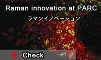
"A team of researchers led by FUJITA Katsumasa (Professor, Graduate School of Engineering), KAWATA Satoshi (Professor, Graduate School of Engineering), and HASHIMOTO Hitoshi (Professor, Graduate School of Pharmaceutical Sciences) at Osaka University succeeded in nearly doubling the conventional resolution of Raman scattering microscopy, which is used in a wide variety of fields in science and industry ranging from drug discovery to device development. Raman scattering is a very subtle effect, so it was thought difficult to increase the resolution of Raman scattering microscopy. However, this group succeeded in measuring at high spatial resolution by shedding dashed light on samples, achieving the highest resolution of microscopy using propagating light. This group’s research results have made it possible to observe spatial distribution of drugs and device materials in further detail, which is expected to be helpful in enhancing the quality and functionality of products." (ResOU, Osaka University.)  The research paper entitled “Structured line illumination Raman microscopy” appeared at the Nature Physics website (Published 2 Dec 2015). The research paper entitled “Structured line illumination Raman microscopy” appeared at the Nature Physics website (Published 2 Dec 2015).
Raman images of a mouse brain slice. (a,b) The intensity distribution of Raman peaks at 1,682 cm.1 (red) and 2,848 cm.1 (green) imaged by the line illumination (LI) (a) and structured-line illumination (SLI) (b) microscopes (Scale bar, 5 mm).
|
|
|
|
|
|


|
|